Our ChromaLIVE™ non-toxic dye products

A Data-Rich Dye
ChromaLIVE™: truly non-toxic for the highest-fidelity biological data
Although profiling drugs with images is a powerful and scalable approach, a fundamental problem persists: are we really probing the true biological response?
Images provide both overlapping and unique phenotypic data compared to omics, but fixation, washing, and toxic probes directly affect cells and the very features used to build those powerful image-based phenotypic profiles.
ChromaLIVE™ is the leading technology for high-throughput phenotypic profiling, enabling researchers to generate richer, more biologically relevant profiles than traditional fixed-cell approaches, all while maintaining similar throughput.
The non-toxicity of ChromaLIVE™ is a unique feature not present with alternative "live-cell dyes" or live cell painting kits, which is crucial to allow accurate phenotypic profiling in live cells, and new kinetic insights.
ChromaLIVE™ Features
Data-rich dye for new cell insights
Non-Toxic
Unaffected gene expression patterns and stable in multi-week live cell cultures. Ideal for downstream assays.
Mix-and-read
No wash, no fixation, no plate copies. With its one-step protocol, it's easy to use ChromaLIVE™. It does not fluoresce in media, removing the need for a washing step.
Data-rich
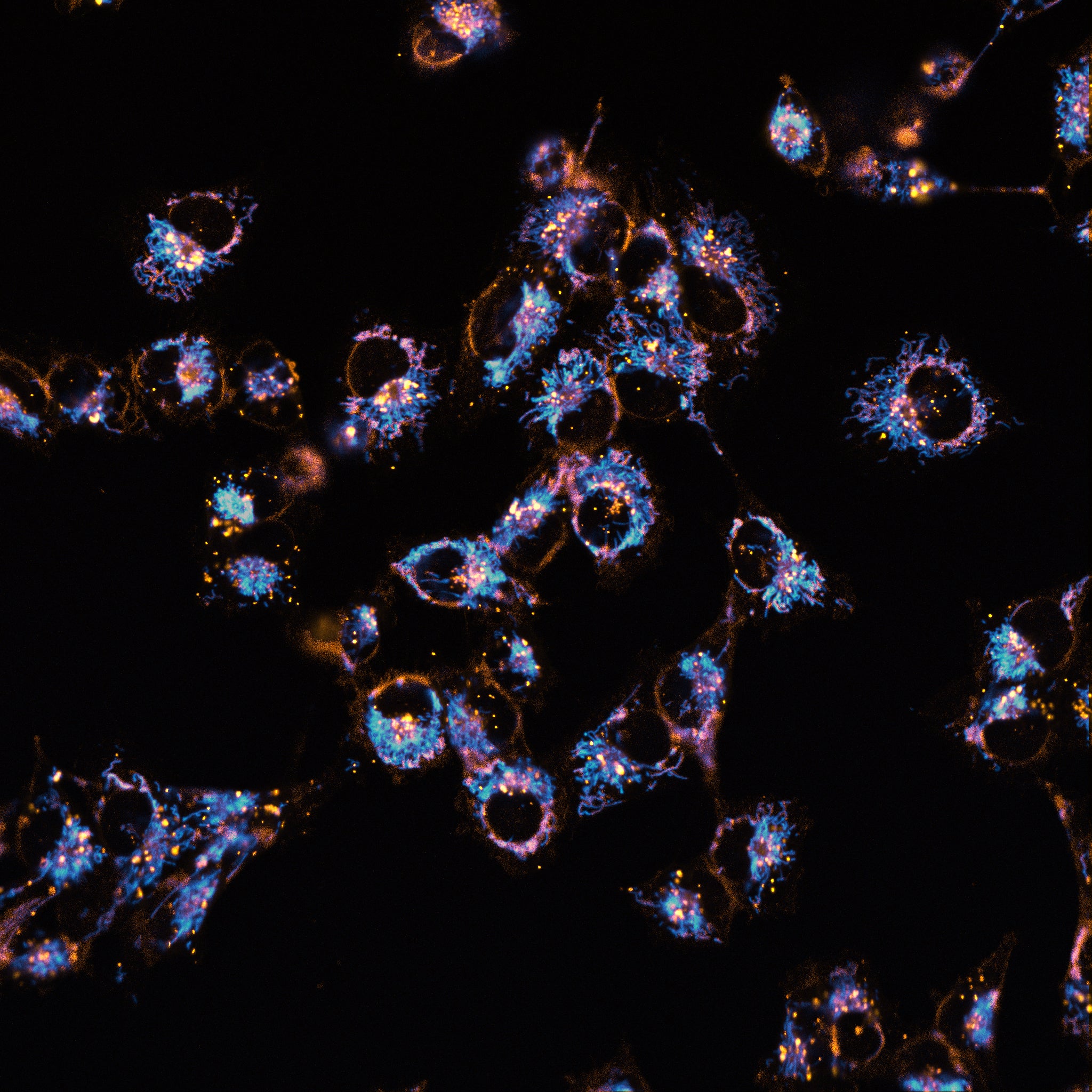
ChromaLIVE™ is a multi-chromatic dye that provides high-density biological information, and therefore helps differentiate subtle phenotypic signatures.


Applications
Richer Data and New Insights Hidden in Cell Physiology that is Unperturbed

Live Cell Painting
Capture data that others omics can't, and go beyond cell painting with the highest-quality cell imaging data. Differentiate diseased and healthy phenotypes, and hint at drug mechanism or off-target effects in high-throughput. Capture valuable insights hidden in the temporal dimension.

AI Models Fed with the Highest Quality Data
Because Saguaro dyes are non-toxic, drug discovery scientists and cell biology researchers can get the most accurate picture of cell response, and the highest-quality cell data at scale to train their powerful AI models.

Organoids and iPSCs
Saguaro dyes and ChromaLIVE™ provide unequaled penetration of 3D cultures, and they are perfect for sensitive cell systems. Get uniform staining of 3D cultures, or differentiate between key steps in the differentiation process of stem cells.

ChromaLIVE™+ Lipo
We are further expanding our ChromaLIVE+™ portfolio with
ChromaLIVE+™ Lipo, a complementary addition to our phenotypic profiling offering. By combining our non-toxic LipoLIVE™ dye with the ChromaLIVE™ dye, additional layers of biologically relevant information can be captured in live-cell systems. ChromaLIVE+™ Lipo enhances high-content imaging workflows by supporting improved bioactivity detection and increased phenotypic distinctiveness.

ChromaLIVE+™ ER
Building on our ChromaLIVE technology, ChromaLIVE+™ ER pushes the boundaries of image-based profiling even further. The combination of our new, non-toxic ER-LIVE™ dye and the ChromaLIVE™ dye enables the capture of additional, relevant biological information. This leads to increased phenotypic profiling performance and enhanced interpretability. ChromaLIVE+™ ER is now the industry-leading solution for profiling cellular responses using high-content images, delivering higher bioactivity detection and phenotypic distinctiveness.

ChromaLIVE™ Deep Red
ChromaLIVE™ Deep Red is just like the original ChromaLIVE™ : same biological inertness and same leading technology for drug profiling, but with a different fluorescence spectrum. This means that ChromaLIVE™ Deep Red can be used with NucleoLIVE™ non-toxic nuclear dye to get even more insight from your live cell painting assays. See the ChromaLIVE™ Deep Red protocol and technical note for more information.

ChromaLIVE™
ChromaLIVE™ is our original non-toxic dye for live-cell imaging and phenotypic profiling. It enables image-based analysis of cells in their native environment, without fixation or toxicity. This multi-chromatic dye provides high-density biological information and helps differentiate subtle phenotypic signatures. ChromaLIVE™ can be used with NucleoLIVE™ Blue non-toxic nuclear dye.
Check Out Our Resources


Frequently Asked Questions
-
RNA sequencing data and week-long cell cultures support this claim. Figure 2 of this SLAS Discovery article presents supporting RNA sequencing and proteomics data showing unperturbed expression patterns – in contrast with alternative live cell dyes – for cells cultured in presence of ChromaLIVE.
In addition, ChromaLIVE has been successfully tested in sensitive cell systems without affecting cell health for weeks on end. For instance, it has been used in a 3-week culture of iPSC-derived neurons, and a 6-week culture of patient-derived prostate cancer organoids.
-
Yes! ChromaLIVE Deep Red is our only other available color option for now. It is just like the original ChromaLIVE: same biological inertness and same leading technology for drug profiling, but with a different fluorescence spectrum that makes it compatible with NucleoLIVE to get even more insight from your live cell painting assays. See the ChromaLIVE Deep Red protocol and technical note for more information.
-
ChromaLIVE is stable for a very long time in culture - i.e. weeks.
Under normal exposure to the microscope’s light source in live cell experiments, the fluorescence intensity of ChromaLIVE remains constant over those long periods of time. In the unwanted case where live cells stained with ChromaLIVE would be exposed to an abnormally high amount of light, cell health is likely to be significantly affected before even observing signs of unstable fluorescence signal from ChromaLIVE.
That’s because the way ChromaLIVE is used, with its constant presence in excess in culture medium, means that any fluorescent ChromaLIVE molecule that would have undergone photo-bleaching in cells would be replaced by a molecule present in the medium. As a reminder, ChromaLIVE fluoresces only upon incorporation into cells, and remains non-fluorescent in the medium.
-
ChromaLIVE™ is a wash-free, multi-chromatic small molecule dye that is excited at two different wavelengths: 488nm, with a long-stokes shift emission in the red (making it compatible with your own GFP fluorophore) and a 561nm excitation (or 640nm excitation for our ChromaLIVE™ Deep Red).
See our document “How does ChromaLIVE™ work” in our resources for more details such as how it stains multiple cell compartments at the same time: https://www.saguarobio.com/resources/
-
Fluorescence signal from ChromaLIVE can both be retained or removed after fixation, depending on your downstream assay needs. Signal retention requires our ChromaLIVE Fix additive and is performed with 4% PFA. By fixing and permabilizing cells (without ChromaLIVE Fix additive) signal can be entirely removed.
-
ChromaLIVE is multichromatic and is therefore excited at two wavelengths: 488nm and 561nm. The 488 channel has a long-stokes shift emission in the red (making it compatible with your own GFP fluorophore), while the 561 channel has standard yellow emission. Most high-content imaging instruments are compatible with ChromaLIVE. Refer to our resources on our website to find a list of compatible instruments, along with technical notes detailing filter selection specific to the Yokogawa, Molecular Devices and Revvity systems. Incucyte systems are also compatible with ChromaLIVE!
-
ChromaLIVE comes in tubes of 10uL, and is used at a dilution of 1:1,000 in culture medium. The biological inertness of ChromaLIVE and its fluorescence only upon incorporation into cells allow its addition at the cell seeding stage, which is highly recommended. Addition of ChromaLIVE at a later stage is possible, but it is recommended to test staining kinetics as optimal staining can take up to 12 hours depending on cell type.
-
Reach out for a quote! Volume pricing is available, and academic discounts too.
Interested in our live cell dyes?
Join the 200+ drug discovery groups relying on Saguaro dyes for better drug insights